Tiếng Việt


Laboratory Equipment


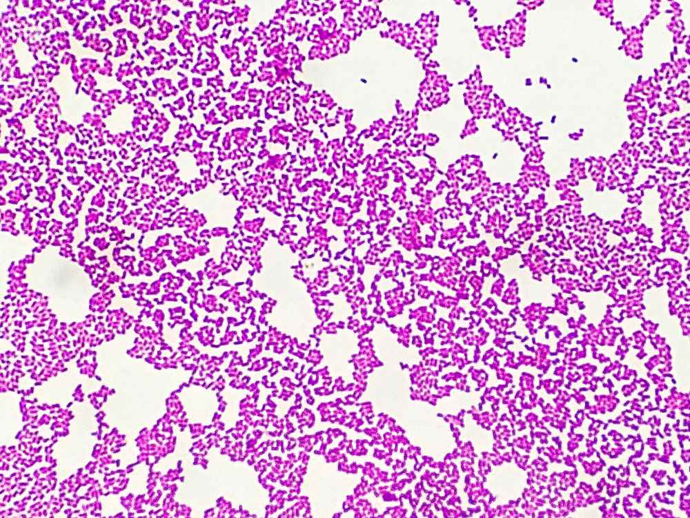
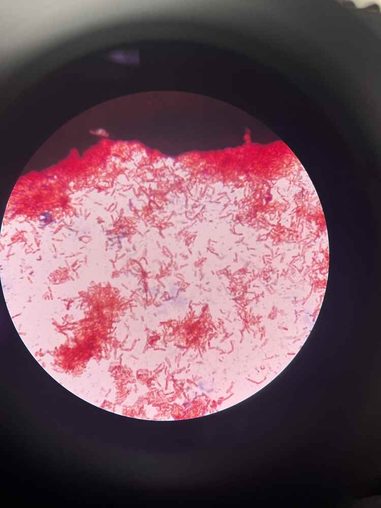
Gram-negative bacilli are rod-shaped bacteria (straight or slightly curved) that belong to the Gram-negative group according to the Gram staining classification. When stained using the Gram method, they appear pink or red under the microscope.
Gram-negative bacilli are widely present in natural environments such as soil, water, the human and animal digestive systems, as well as in hospital settings. They can become pathogenic agents when the body’s immune system is weakened or when favorable conditions exist, such as open wounds, invasive medical procedures, or poor hygiene.
During staining, Gram-negative bacteria do not retain the crystal violet purple color; they are decolorized by alcohol and then take up the red or pink color from safranin. This characteristic reflects a thinner cell wall with less peptidoglycan and an outer membrane containing lipopolysaccharide (also called endotoxin), which triggers strong inflammatory responses in the human body.
Typically belong to the Enterobacteriaceae family, capable of fermenting glucose to produce energy.
Typical representatives include
Escherichia coli coliform bacillus, Klebsiella pneumoniae pneumonia bacillus, Salmonella typhoid bacillus, Shigella bacillary dysentery. This group commonly causes infections in the gastrointestinal tract, urinary tract, and bloodstream.
They do not ferment glucose but only oxidize it. Common types include Pseudomonas aeruginosa (blue-green pus bacillus), Acinetobacter baumannii, Burkholderia cepacia, Stenotrophomonas maltophilia.
This group often causes hospital-acquired infections, especially in immunocompromised patients, with an isolation rate of approximately 12 percent among all pathogenic bacteria according to recent studies.
Gram-negative bacilli are not only pathogenic bacteria; some are also normal components of the microbiota in the body, such as in the intestines or female genital tract. However, when imbalance occurs, they can lead to serious infections.
The cell structure of Gram-negative bacilli is quite complex and different from Gram-positive bacteria, contributing to their antibiotic resistance and pathogenicity. Specifically:
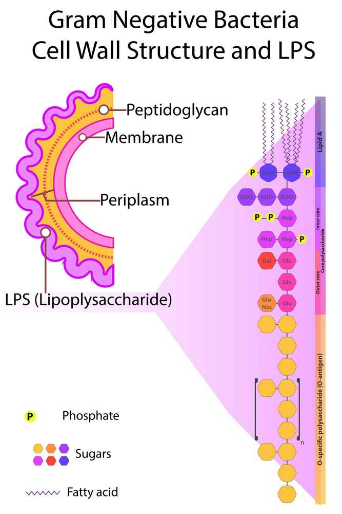
This structure makes Gram-negative bacilli resistant to penicillin due to thin peptidoglycan but sensitive to some other antibiotics, while the endotoxin causes strong inflammatory responses.

There are hundreds of types of Gram-negative bacilli, but the most common include:
These bacteria are commonly found in the environment and become dangerous when they enter through skin, mucous membranes, respiratory, or digestive routes.
Gram staining is an important differential staining technique in microbiology. Originally, Gram developed this method to differentiate pneumonia-causing bacteria in lung tissue, but today it remains the fastest, simplest, and most essential preliminary identification method in clinical microbiology. This technique uses two contrasting dyes—crystal violet and safranin—to classify bacteria into two main groups:
Gram-positive and Gram-negative based on cell wall structure. Gram staining allows rapid diagnosis in just a few minutes, supporting initial antibiotic selection and guiding further tests. However, the method can be affected by factors such as bacterial age (use fresh, actively growing samples), smear thickness, decolorization time, or technical errors (e.g., over-decolorization can cause false Gram-negative appearance in Gram-positive bacteria).
See also: What are Gram-positive bacilli? Structure and classification?
Gram-negative bacilli are a diverse group that accounts for a large proportion of community-acquired and hospital-acquired infections. They have a more complex structure than Gram-positive bacteria, with an outer LPS membrane causing high toxicity, leading to strong immune responses and common antibiotic resistance. Gram staining plays a key role in early detection, helping to classify and select appropriate antibiotics.

Gram staining directly from clinical specimens (sputum, urine, pus, blood, cerebrospinal fluid) helps quickly detect rod shape and Gram-negative pink/red color, supporting initial diagnosis.
Gram-negative bacilli are not only rod-shaped bacteria but also represent the complexity of prokaryotic cell structure, with thin peptidoglycan and an outer LPS membrane that causes them to take up pink color in Gram staining. This explains their strong pathogenicity, difficulty in treatment, and the need for rapid diagnosis. Understanding the mechanism helps clinicians make accurate diagnoses, select appropriate antibiotics, and control infections more effectively, reducing mortality rates.