Tiếng Việt


Laboratory Equipment


Gram stain is one of the most important and widely used staining techniques in microbiology. Developed by Danish scientist Hans Christian Gram in 1884, this method allows scientists and clinicians to classify bacteria into two major groups based on the chemical and physical properties of their cell walls. Even after more than a century, gram stain remains a foundational tool in clinical diagnostics, research laboratories, and infection control programs around the world.
Understanding the gram stain procedure, gram stain results interpretation, and the difference between gram positive and gram negative bacteria is essential for anyone studying microbiology, working in a clinical laboratory, or pursuing a career in medicine, nursing, pharmacy, or biomedical science. This comprehensive guide explains what gram stain is, how to perform gram stain correctly, why it matters, and how results are interpreted in real world settings.
Gram stain is a differential staining technique used to distinguish between two broad categories of bacteria: gram positive and gram negative. The technique relies on differences in the structure of bacterial cell walls, particularly the thickness of the peptidoglycan layer. Because these structural differences affect how bacteria retain or release certain dyes, gram stain provides a quick and reliable way to categorize unknown bacterial samples.
In simple terms, gram stain answers one basic question: does the bacterial cell wall retain a violet dye after exposure to a decolorizing agent, or does it lose that dye and instead take up a pink or red counterstain? The answer to that question places the organism into one of two categories, each with distinct clinical and biological implications.
Hans Christian Gram originally developed this staining technique while working with lung tissue samples from patients who had died of pneumonia. His goal was to better visualize bacteria under a microscope so that pathogens could be distinguished from surrounding human tissue. Although Gram himself did not fully understand why some bacteria stained differently than others, his technique proved remarkably effective and has stood the test of time.
Over the decades, researchers discovered that the underlying reason for the staining difference lies in the composition of the bacterial cell envelope. This discovery transformed gram stain from a simple visualization tool into a technique with deep biological meaning, one that reflects fundamental differences in bacterial physiology.
To understand why the gram staining technique works, it helps to look closely at bacterial cell wall anatomy.
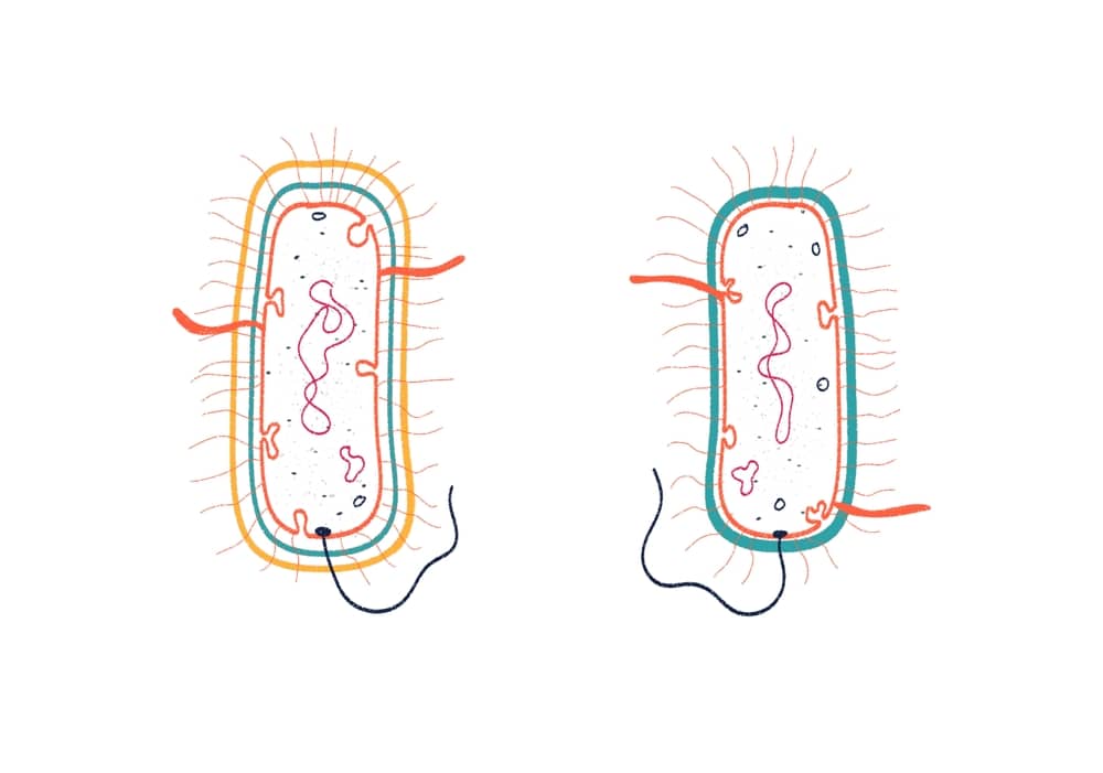
Gram positive bacteria have a thick layer of peptidoglycan, sometimes composed of dozens of layers, located just outside their plasma membrane. This thick mesh like structure is able to trap the crystal violet iodine complex formed during the staining process. As a result, gram positive bacteria retain the purple color even after the decolorization step.
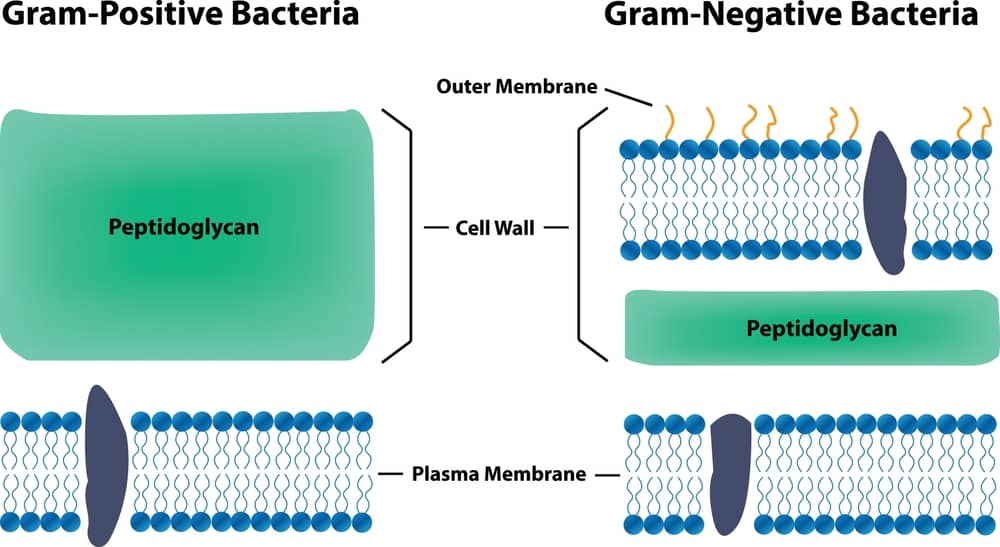
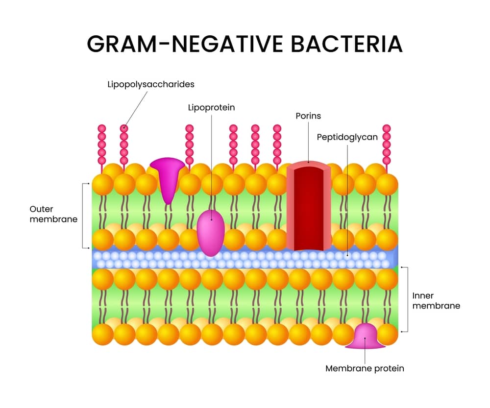
Gram negative bacteria have a much thinner peptidoglycan layer, and importantly, they possess an additional outer membrane composed of lipopolysaccharides and phospholipids. During the decolorization step of the staining procedure, alcohol or acetone disrupts this outer membrane and washes away the crystal violet iodine complex from the thin peptidoglycan layer. Because the purple dye is lost, gram negative bacteria appear colorless until a counterstain, typically safranin, is applied to give them a characteristic pink or red appearance.
A standard gram stain kit contains four reagents, applied in a fixed order. Using correctly prepared reagents is just as important as following the correct timing.
| Reagent | Typical Composition | Approximate Contact Time | Purpose |
|---|---|---|---|
| Crystal violet | 2% crystal violet in ethanol, mixed with 1% ammonium oxalate | 30 to 60 seconds | Primary stain, colors all cells purple |
| Gram iodine | 1% iodine and 2% potassium iodide in water | 30 to 60 seconds | Mordant, forms a large dye complex inside the cell |
| Decolorizer | 95% ethanol, or a 1 to 1 acetone alcohol mixture | 3 to 10 seconds, added dropwise until runoff is clear | Removes dye from thin walled cells only |
| Safranin | 0.25% safranin O in water or dilute ethanol | 30 to 60 seconds | Counterstain, colors decolorized cells pink or red |

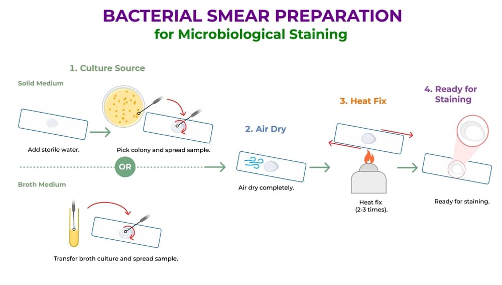
Performing a gram stain involves several carefully timed steps. While exact protocols can vary slightly between laboratories, the general gram stain procedure includes the following stages.

Even experienced technicians can introduce error into a gram stain. The most frequent mistakes include:
| Problem | Likely Cause | Appearance on Slide | Correction |
|---|---|---|---|
| Over decolorized | Decolorizer left on too long, or applied in excess | Gram positive bacteria falsely appear pink or red, mimicking gram negative organisms | Reduce decolorizer contact time; stop as soon as runoff turns clear, not colorless for an extended period |
| Under decolorized | Decolorizer removed too quickly or applied unevenly | Gram negative bacteria falsely retain purple color, mimicking gram positive organisms | Apply decolorizer drop by drop until the runoff is visibly clear before moving to the counterstain |
| Smear too thick | Excess sample applied to the slide | Cells appear uniformly dark or purple regardless of true gram reaction | Dilute the sample and prepare a thinner, more even smear |
| Old culture artifact | Culture older than 24 hours | Gram positive organisms may appear gram variable, showing a mix of purple and pink cells | Use fresh, actively growing cultures whenever possible |
| Weak or expired reagents | Reagents stored too long or contaminated | Faint or inconsistent staining across the entire slide | Replace reagents on a regular schedule and always run a control slide |
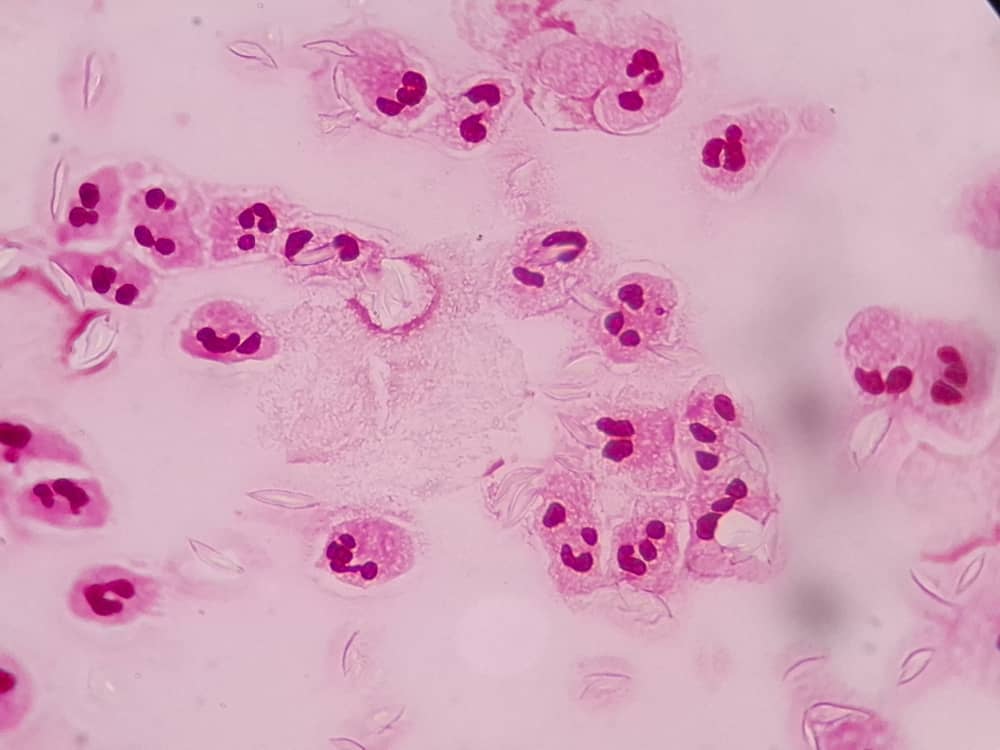
Once the staining process is complete, gram stain results interpretation focuses on three main characteristics: color, shape, and arrangement.
Bacteria generally fall into a few basic shapes, including:
The way bacterial cells group together also provides useful clues about species identity. Common arrangements include clusters, chains, pairs, and tetrads.
Combining color, shape, and arrangement allows laboratory professionals to make an educated preliminary identification, such as gram positive cocci in clusters, which often suggests Staphylococcus species, or gram negative rods, which may point toward organisms such as Escherichia coli or Pseudomonas aeruginosa.

Not every bacterium responds predictably to the gram staining technique. Organisms in the Mycobacterium genus, such as those causing tuberculosis, have a waxy, lipid rich cell wall containing mycolic acids that resists both crystal violet uptake and standard decolorization. These organisms require a separate method called acid fast staining, most commonly the Ziehl Neelsen or Kinyoun technique.
| Feature | Gram Stain | Acid Fast Stain |
|---|---|---|
| Target organisms | Most bacteria | Mycobacterium species and a few related organisms |
| Primary dye | Crystal violet | Carbol fuchsin |
| Key differentiator | Peptidoglycan thickness | Mycolic acid content in the cell wall |
| Heat required | No | Yes, heat is typically used to drive dye into the waxy wall |
| Typical clinical use | General bacterial identification | Diagnosis of tuberculosis and related mycobacterial infections |
Because a standard gram stain often shows Mycobacterium species as faint, poorly stained, or “gram ghost” cells rather than a clear positive or negative reaction, clinicians who suspect tuberculosis or a related infection will order acid fast staining in addition to, or instead of, a routine gram stain.
Gram stain plays a central role in diagnosing infections and guiding treatment decisions. Its importance can be understood through several key applications.
Culture based identification of bacteria can take twenty four to forty eight hours or longer. Gram stain, on the other hand, can be performed and interpreted within minutes. This speed allows physicians to begin an educated guess about the likely category of pathogen almost immediately, which is especially valuable in cases of severe infection such as sepsis or meningitis.
Because gram positive and gram negative bacteria often respond differently to various classes of antibiotics, knowing the gram reaction of an infecting organism helps clinicians choose an appropriate empiric antibiotic before final culture and sensitivity results become available. For more background on how this choice is later confirmed, see antibiotic susceptibility testing.
Gram stain is also used to verify the purity of bacterial cultures grown in the laboratory. If a culture that should contain a single organism shows more than one distinct morphology or gram reaction under the microscope, this may indicate contamination.
In some clinical scenarios, repeated gram stains of body fluids, wound samples, or respiratory secretions can help track whether an infection is resolving or worsening over time.
Cerebrospinal fluid sample.
A patient presents with fever, neck stiffness, and altered mental status. A gram stain of cerebrospinal fluid shows gram positive diplococci, prompting immediate empiric therapy for suspected pneumococcal meningitis while culture results are pending.
Sputum sample.
A hospitalized patient with a productive cough and new fever has a sputum gram stain performed. The slide reveals numerous gram negative rods along with many white blood cells, guiding the care team toward coverage for gram negative respiratory pathogens such as Klebsiella pneumoniae.
Wound sample.
A surgical site infection is swabbed, and the gram stain reveals gram positive cocci in clusters. This finding raises early suspicion for Staphylococcus aureus, including the possibility of a resistant strain, and helps direct initial wound care and antibiotic selection.
These examples illustrate why gram stain is often called a bridge between bedside assessment and definitive laboratory identification: it narrows the diagnostic possibilities long before final culture results are available.
Examples of Gram Positive Bacteria
Examples of Gram Negative Bacteria
Limitations of Gram Stain
While gram stain is extremely useful, it does have limitations that laboratory professionals must keep in mind.
Laboratories aiming for consistent and reliable gram stain results should follow several best practices.
Despite the rise of advanced molecular diagnostic tools such as polymerase chain reaction testing and mass spectrometry based identification systems, gram stain remains a cornerstone technique in clinical microbiology laboratories worldwide. Its low cost, speed, and simplicity make it an irreplaceable first step in the identification workflow, often performed before more sophisticated and expensive testing methods, including bacterial cell wall based diagnostics, are pursued.

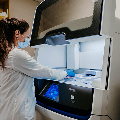
Automated staining machines have also been introduced in many high volume laboratories, helping standardize the process and reduce variability caused by manual technique. However, human interpretation under the microscope remains essential, since experienced microbiologists can recognize subtle morphological clues that automated systems may miss.
According to the Centers for Disease Control and Prevention, rapid diagnostic methods including gram stain continue to play a supporting role in antimicrobial stewardship programs by helping clinicians select more targeted therapy sooner. The American Society for Microbiology also maintains detailed laboratory protocols confirming gram stain as a core competency for clinical microbiology training. For broader international laboratory standards, the World Health Organization publishes guidance on basic bacteriology procedures used in health systems worldwide.
What does a gram stain test for?
Gram stain tests for the presence and classification of bacteria in a clinical or laboratory sample, determining whether organisms are gram positive or gram negative based on their cell wall structure.
How long does a gram stain take?
The staining procedure itself typically takes only a few minutes to complete, and results can usually be interpreted under a microscope within fifteen to thirty minutes of sample collection.
Can viruses be seen with gram stain?
No. Gram stain is designed specifically for bacteria and cannot be used to visualize viruses, which are far too small to be seen with standard light microscopy.
Why do some bacteria not gram stain well?
Certain bacteria have unusual cell wall compositions, such as the waxy, lipid rich walls found in Mycobacterium species, which prevent them from taking up the gram stain dyes effectively and instead require acid fast staining.
Is gram stain still relevant today?
Yes. Even with modern molecular diagnostic advances, gram stain remains a fast, inexpensive, and clinically valuable first line tool for identifying the general category of bacteria present in a sample.
Gram stain continues to be one of the most valuable and widely practiced techniques in microbiology, more than a century after its discovery. By distinguishing gram positive from gram negative bacteria, this simple yet elegant gram staining technique provides critical information that guides diagnosis, treatment, and laboratory quality control. Whether used in a hospital laboratory, a research setting, or an educational classroom, gram stain remains an essential skill for anyone working in the field of microbiology. Mastering the gram stain procedure, understanding its underlying science, recognizing common mistakes, and knowing its limitations are all key steps toward using this technique effectively and confidently in real world applications.
Laboratories that want to trial high quality staining reagents before committing to a full purchase can take advantage of a free gram stain kit placement program offered by Lintech, which allows facilities to evaluate reagent performance under real world testing conditions.